1. Complete Activity 5.1 (Page 49).
• Let us take a small piece from an onion bulb. With the help of a pair of forceps, we can peel off the skin (called epidermis) from the concave side (inner layer) of the onion. This layer can be put immediately in a watch-glass containing water. This will prevent the peel from getting folded or getting dry. What do we do with this peel?
• Let us take a glass slide, put a drop of water on it and transfer a small piece of the peel from the watch glass to the slide. Make sure that the peel is perfectly flat on the slide. A thin camel hair paintbrush might be necessary to help transfer the peel. Now we put a drop of safranin solution on this piece followed by a cover slip. Take care to avoid air bubbles while putting the cover slip with the help of a mounting needle. Ask your teacher for help. We have prepared a temporary mount of onion peel. We can observe this slide under low power followed by high powers of a compound microscope.
What do we observe as we look through the lens? Can we draw the structures that we are able to see through the microscope, on an observation sheet? Does it look like Fig. 5.2?
We can try preparing temporary mounts of peels of onions of different sizes. What do we observe? Do we see similar structures or different structures?
Answer:
Aim: To conclude based on what we see through the microscope.
Materials Required: Onion bulb, forceps, watch-glass, water, glass slide, water, camel hair paintbrush, safranin solution, cover slip, mounting needle, compound microscope.
Procedure:
(i) Using the pairs of forceps, the epidermis from the concave side of the onion is peeled off.
(ii) The layer is put immediately in a watch-glass containing water to prevent folding and drying of the peel.
(iii) Now a glass slide is taken and a drop of water is put on it.
(iv) A small piece of the peel is transferred from the watch-glass to the slide using the thin camel hair paintbrush, making sure that the peel is perfectly flat on the slide.
(v) A drop of safranin solution is put on the piece.
(vi) Then a cover slip is put on it with the help of a mounting needle, taking care to avoid air bubbles. This can be done under teacher supervision.
(vii) Now the slide is observed under low power followed by high powers of a compound microscope. The observations are noted.
(viii) The structures observed are drawn on an observation sheet.
(ix) The activity is repeated using onion peels of different sizes and the observations are noted.
Observations:
- The following structures are seen:
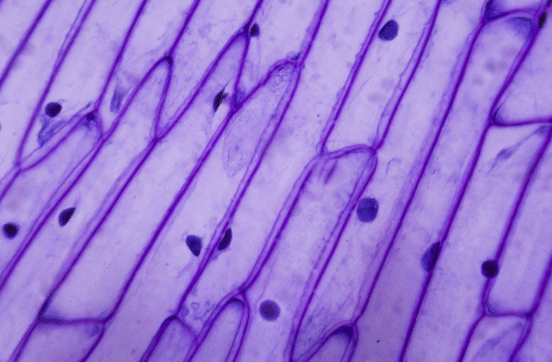
- We see similar structures for onion peels of different sizes.
Conclusions:
- All onion cells are rectangular in shape and have a round nucleus at the centre.
- Together the cells form a big structure called an onion bulb.
- The cells of the onion peel will all look the same, regardless of the size of the onion they came from.
“• Let us take a small piece from an onion bulb. With the help of a pair of forceps, we can peel off the skin (called epidermis) from the concave side (inner layer) of the onion. This layer can be put immediately in a watch-glass containing water. This will prevent the peel from getting folded or getting dry. What do we do with this peel?
• Let us take a glass slide, put a drop of water on it and transfer a small piece of the peel from the watch glass to the slide. Make sure that the peel is perfectly flat on the slide. A thin camel hair paintbrush might be necessary to help transfer the peel. Now we put a drop of safranin solution on this piece followed by a cover slip. Take care to avoid air bubbles while putting the cover slip with the help of a mounting needle. Ask your teacher for help. We have prepared a temporary mount of onion peel. We can observe this slide under low power followed by high powers of a compound microscope.
What do we observe as we look through the lens? Can we draw the structures that we are able to see through the microscope, on an observation sheet? Does it look like Fig. 5.2?
We can try preparing temporary mounts of peels of onions of different sizes. What do we observe? Do we see similar structures or different structures?” – Solved.
Related Links:
Solution to Activity 5.1
Solution to Activity 5.2
Solution to Activity 5.3
Solution to Activity 5.4
Solution to Activity 5.5
Solution to Activity 5.6
Solution to Activity 5.7
Solution to Chapter 5 The Fundamental Unit of Life